A spectrum for circularity
Michelle Sestak at HORIBA North America, explains why spectroscopic microscopy matters in a circular economy.
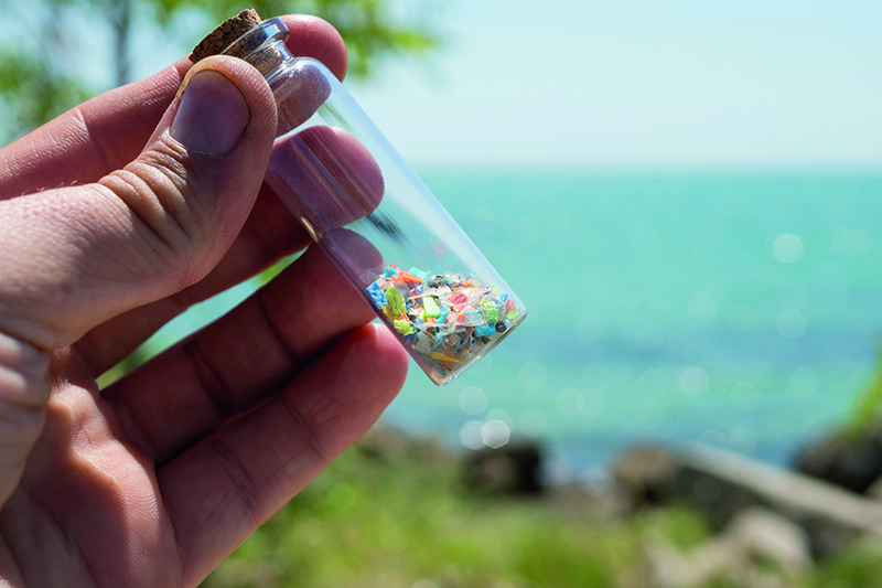
Microplastic fragments collected from a shoreline
© SIVStockStudio / ShutterstockThe transition from a linear economic model to a circular one is arguably the greatest materials science challenge of our generation. For decades, the global supply chain operated on a simple directive – to take, make and dispose of materials that were engineered for performance and durability with scant regard for after they stopped being useful. Now, the paradigm has shifted. We are tasked with recovering, recycling and remanufacturing materials to keep them in an endless loop of utility.
However, a fundamental problem lies at the heart of this transition. You cannot effectively recycle what you cannot accurately identify. The circular economy is heavily reliant on purity. When materials are mixed, contaminated, or chemically degraded, their value plummets. Mechanical sorting facilities do an admirable job of separating bulk items, but the true battle for circularity is fought at the microscopic level.
To truly close the loop on plastics, critical minerals and electronic waste, materials scientists are turning to advanced imaging and spectroscopic microscopy. With techniques such as Raman microscopy, X-ray fluorescence (XRF) imaging and fluorescence spectroscopy, researchers can visualise the chemical and elemental realities of recycled materials. This depth of insight is now an industrial necessity for accurate Life Cycle Assessments (LCAs) and ensuring that recycled materials meet the rigorous performance standards of virgin feedstocks.
The illusion of purity
Plastics present one of the most visible challenges to the circular economy. The modern packaging industry relies heavily on complex and engineered materials. A seemingly simple food wrapper is rarely a single sheet of plastic. It is often a highly engineered multilayer film designed to provide specific barriers against moisture, oxygen and light. A single film might contain layers of polyethylene, polypropylene, ethylene vinyl alcohol and various adhesive tie layers.
When this multilayer film enters a mechanical recycling stream, it becomes a nightmare. Melted together, these incompatible polymers form a low-value blend with poor mechanical properties, often downcycled to park benches or speed bumps. To achieve true circularity and turn old food packaging into new food packaging, chemical recycling and advanced compatibilisation techniques are required. But first, engineers must know exactly what they are dealing with.
This is where Raman microscopy becomes an invaluable tool. Raman spectroscopy relies on the inelastic scattering of light. When a laser illuminates a sample, most of the light scatters at the exact same wavelength as the laser source. However, a tiny fraction of the light interacts with the material’s molecular vibrations, shifting in energy. This creates a unique spectral fingerprint that can definitively identify the molecule’s chemical structure.
When this spectroscopic technique is coupled with an optical microscope, it transforms from a bulk analysis tool into a high-resolution chemical imaging system. Confocal Raman microscopy allows researchers to focus the laser into a tiny volume, often smaller than a cubic micrometre. By scanning the laser across a sample, the instrument collects thousands of individual spectra, building a comprehensive and colour-coded chemical map of the material.
Bringing it to light
Consider the analysis of a recycled, multilayer, polymer pellet. Using conventional thermal analysis, a scientist might be able to tell that the pellet contains both polyethylene and polypropylene, but it cannot reveal how those polymers are distributed. Are they perfectly homogenised, or are there distinct domains of incompatible material?
Using a Raman microscope, a researcher can slice the pellet, polish the cross-section and map the surface. The resulting image shows a sample’s physical topography and the exact spatial distribution of the chemical components. The software assigns a colour to the unique Raman signature of polyethylene and a different colour to polypropylene. If the recycling process involves a compatibiliser to help these polymers blend, the Raman map will visually confirm whether the compatibiliser successfully dispersed the polymer domains, or if large, failure-prone clumps remain.
In the context of designing new, recyclable-by-design packaging, manufacturers are using Raman imaging to measure the exact thickness of microscopic tie layers in newly extruded films. If a barrier layer is too thin, the product will spoil. If it is too thick, it wastes material and complicates the recycling process. Raman microscopy provides the non-destructive, sub-micrometre chemical vision required to optimise these precise material architectures.
The microplastic conundrum
The drive towards a circular economy has an unintended consequence. The very act of mechanically shredding, washing and extruding recycled plastics generates a significant amount of microplastics. These tiny fragments of polymer, typically defined as being <5mm in length, can easily escape recycling facilities and enter the industrial wastewater stream.
Tracking and mitigating these microplastics requires specialised analytical techniques. Standard optical microscopy can count particles, but it cannot distinguish among harmless organic debris, such as a grain of sand and a fragment of hazardous polystyrene. Automated Raman microscopy offers a robust solution for environmental monitoring within recycling plants.
Water samples from the recycling facility can be filtered through a fine mesh, trapping the suspended particles. The filter is then placed under the Raman microscope. Advanced software systems automatically locate every single particle on the filter, determine its size and shape, and then fire the laser to acquire a chemical spectrum. The system rapidly compares the collected spectra against a vast library of known polymers.
Within a few hours, a plant manager can receive a detailed report outlining not only the total number of particles in the water, but the exact chemical makeup of each one. If a sudden spike of PET microparticles is detected, operators can trace the issue back to a specific shredding or washing step in the plant, optimising the machinery to reduce environmental leakage.
The battery boom
While plastics dominate the consumer waste conversation, the rapid electrification of the automotive sector has introduced an entirely different circularity challenge. Millions of lithium-ion batteries will reach the end of their useful lives over the next decade. These batteries contain critical and expensive minerals, including cobalt, nickel, manganese and lithium. Recovering these metals through battery recycling is a global priority.
The initial stage of battery recycling involves shredding the depleted cells to create a powdery mixture known as black mass. This black mass is a chaotic blend of cathode materials, graphite from the anode, aluminium and copper current collectors, and degraded electrolyte. To refine this black mass into high-purity precursors for new batteries, hydrometallurgical processing is required.
Plant operators must know the elemental composition of the black mass batch to calculate the correct dosage of acids and solvents for extraction. While techniques like inductively coupled plasma optical emission spectroscopy detect light elements such as lithium, XRF microscopy provides the necessary elemental vision for heavier transition metals.
When a sample is bombarded with high-energy X-rays, the inner shell electrons of the atoms are ejected. As outer shell electrons drop down to fill the vacancies, they emit secondary fluorescent X-rays. The energy of these emitted X-rays is strictly characteristic of the element that produced them.
Micro-XRF instruments can focus an X-ray beam down to a spot size of just a few micrometres. By mapping a sample of black mass, researchers can see the exact distribution of heavy elements. They can identify large agglomerations of copper that might foul the chemical leaching process. They can map the ratios of nickel, manganese and cobalt to determine the original chemistry of the shredded batteries, which dictates the economic value of the recycled batch.
Furthermore, XRF is highly sensitive to heavy metal contaminants. If a batch of black mass is contaminated with lead or cadmium from improper sorting at the recycling facility, the XRF map will instantly highlight the localised presence of these toxic elements, preventing an entire batch of expensive recycled material from being ruined.

Service technician repairs electric battery pack of a hybrid car
© Scharfsinn / ShutterstockTotal characterisation
The true power of modern materials analysis lies in correlative microscopy. This approach involves analysing the same microscopic region of a sample using two or more different techniques to build a complete picture of its physical, chemical and elemental state. In the context of battery recycling, combining XRF with Raman microscopy offers profound insights.
Suppose a researcher is investigating why a specific batch of recycled cathode material is underperforming in test cells. First, they use micro-XRF to map the elemental distribution. The XRF map confirms that the ratio of nickel, manganese and cobalt is perfectly uniform. The elemental purity is excellent.
Next, they transfer the sample to a Raman microscope, using software to locate the exact coordinates mapped by XRF. While XRF detects the elements, Raman detects the molecular bonds and crystalline structure. The Raman spectra reveal that the oxygen bonds within the metal oxide lattice have fundamentally shifted. The material has undergone a phase transition during the recycling process, moving from a highly ordered layered structure to a disordered spinel structure.
By correlating the elemental data with the structural data, the researcher deduces that the hydrometallurgical recovery process was too aggressive, damaging the crystal lattice even while maintaining elemental purity. This level of root-cause analysis is impossible with a single technique, proving that advanced imaging is critical for optimising recovery processes.
Industrial water
Circularity is not limited to solid materials. Industrial processes, including recycling, consume vast quantities of water. Achieving a closed-loop system requires recycling the processed water itself, which introduces the challenge of monitoring dissolved contaminants.
Fluorescence spectroscopy plays a vital role in this area. Unlike Raman, which relies on light scattering, fluorescence occurs when a molecule absorbs light of a specific wavelength and then emits light at a longer, lower-energy wavelength. Many organic molecules, including those derived from degraded plastics, industrial solvents and natural organic matter, exhibit strong natural fluorescence.
By using an advanced technique called an excitation-emission matrix, scientists can generate a 3D topographical map of the fluorescent compounds within a water sample. The instrument scans through a wide range of excitation wavelengths while simultaneously measuring the emission spectra. The resulting visual matrix acts as a unique optical fingerprint for the water sample.
In a recycling plant, engineers can take the fingerprint of clean, baseline process water. If a subsequent batch of recycled water shows a new cluster of fluorescent peaks, it immediately indicates that a specific organic contaminant has bypassed the filtration system. This rapid and highly sensitive technique allows facilities to monitor water quality in near real-time, ensuring that process water can be safely reused without introducing invisible contaminants back into the manufacturing loop.
The value of spectral data
The ultimate goal of the circular economy is to reduce the environmental footprint of human industry. To quantify this reduction, organisations rely on LCAs. An LCA attempts to calculate the total environmental impact of a product from raw material extraction through to its end of life.
Historically, LCAs for recycled materials have relied on broad assumptions. An LCA might assume that a mechanical recycling process yields a plastic with 90% of the structural integrity of virgin material. However, these assumptions are often flawed, leading to inaccurate carbon accounting and sub-optimal engineering decisions.
Spectroscopic microscopy replaces these assumptions with hard data. When an engineer uses Raman imaging to prove that a compatibilised recycled polymer blend has achieved complete microscopic homogeneity, that data feeds directly into the LCA. It proves that the recycled material can be used in higher-demand applications, displacing more virgin plastic than previously estimated.
Similarly, when micro-XRF data confirms that a batch of recycled battery precursors contains less than 10ppm of heavy metal contamination, battery manufacturers can confidently incorporate that material into high-performance cells. The empirical test results generated by these imaging techniques provide the foundational trust required for industries to fully embrace recycled feedstocks.
The circular economy will not be built solely on good intentions and broad policy mandates. It will be built by materials scientists and engineers fighting for marginal gains in purity, efficiency and material performance.
We are moving away from a world where materials are simply melted down and remoulded, hoping for the best. We are entering an era of precision recycling. Advanced analytical tools are no longer confined to academic research laboratories. They have become the essential diagnostic tools of the new industrial revolution.
By illuminating the invisible structures, elements and contaminants hidden within our waste streams, these technologies provide the roadmap for true sustainability. They prove that with the right vision, there is no such thing as waste, only materials waiting to be understood, engineered and used once again.
More like this...