Sub-surface sensitivity
Tim Nunney of Thermo Fisher Scientific, USA, analyses the evolution of X-ray photoelectron spectroscopy.
In materials science, the most interesting stories are often written at the surface, although not necessarily the surface that defines the material’s outer boundary. They can quite often lie buried at interfaces, as gradients or hidden layers, where chemistry shifts quietly but decisively to help or hinder the desired performance.
X-ray photoelectron spectroscopy (XPS) is a technique that is valued for its surface sensitivity. It has steadily evolved to meet the challenge of probing beneath the outer layer, extending its reach from the outermost few nanometres into the depths below.
The method of XPS depth profiling involves sequential steps of data collection, followed by removal of material, to move the analysis point from the surface and into the sub-surface region. The spectra collected at each level are then used to create a composition profile. It is an experimental method that has been used for decades, growing from the need to clean the surface for analysis to removing ultra-thin layers of adventitious contamination, and
then reaching several micrometres deep into a sample.
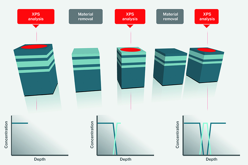
Schematic of XPS depth profiling experiment
© Thermo Fisher ScientificThe process has evolved to reflect the changing nature of the materials being analysed. As spectrometers improved, it became possible to see that, in some cases, the material removal step was altering the sample, and this became a key impetus for advances in depth profiling. Each step forward has expanded capability, but none has rendered its predecessors obsolete. Instead, the field now resembles a carefully assembled toolkit, where the choice of method depends as much on the material as on the question being asked.
In the spotlight
For decades, monatomic ion beams were the only method for material removal during XPS depth profiling experiments. Typically using ions such as argon, these systems remove material through a largely ballistic process.
Energetic ions, with a beam energy in the range of 100-5,000eV, strike the surface, transferring momentum and causing atoms to eject in cascades that gradually carve through layers. The ion beam is essentially rastered across the surface to create an etch crater. This is set to five times the size of the analysis area, ensuring that data is collected from the crater’s flat bottom, preventing degradation in depth resolution caused by edge effects. By controlling ion energy, researchers can tailor removal rates, enabling both delicate surface cleaning and deeper profiling.
This approach has been applied to a wide range of sample types. Passivation coatings on metals, multilayer coatings for glass and semiconductor stacks can all be explored with nanometre-scale precision. In many inorganic systems, particularly metals and stable oxides, monatomic sputtering continues to deliver reliable and reproducible results.
However, monatomic ion beams are not appropriate for all sample types. Polymers experience damage to their chemistry when exposed to even very low-energy monatomic ion beams. Such beams can also change the chemistry of some inorganic materials, such as certain oxides and III/V semiconductors.
As material removal depends on the energy transfer from the impacting ion to the surface, lighter elements, such as oxygen, can be preferentially removed, while heavier species remain. As the number of ion exposures increase, the apparent material composition is altered, causing variation in elemental composition, or inducing chemical changes such as reduction. Therefore, a solution was needed.
A softer approach
To meet these requirements, given the growing development of polymer-based devices, gas cluster ion beams were developed. Rather than individual atoms, singly charged clusters of tens to thousands of atoms are accelerated towards the surface. The total beam energy is distributed across the cluster, reducing the energy per atom.
The effect is a softer interaction between the projectile and the surface. The weakly bound cluster penetrates much less deeply into the surface, limiting the sub-surface collision cascades. This minimises surface modification of the remaining material to a much thinner region, limiting its influence within the XPS analysis depth.
Cluster ion beams have extended XPS depth profiling to encompass materials that were previously inaccessible. Polymers can now be profiled over micrometre depths while retaining key functional groups.
Clusters of 1,000 to 2,000 atoms are commonplace for profiling polymers, but it was soon discovered that using a high beam energy and reducing the cluster size to 300 atoms or less means that inorganic materials could be successfully measured, when previously there was an issue with monatomic ion beams.
However, while cluster beams reduce damage, they do not eliminate all artefacts. Preferential sputtering can still occur within inorganic systems. The impact of the cluster ion at the surface can cause a large local temperature increase, which can cause preferential sputtering if components have different sublimation temperatures. Structural modifications may also be induced that influence the measured composition. Therefore, a new approach was needed to analyse these problem samples.
Rapid fire
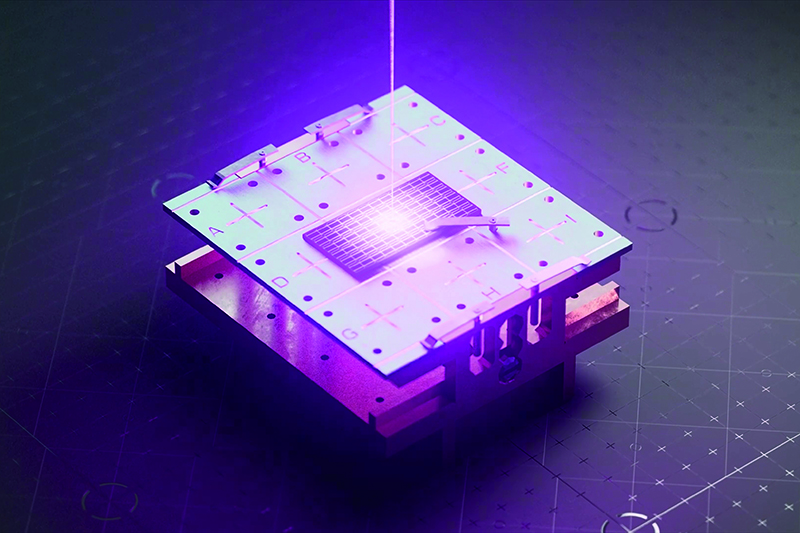
In theory, if a method of material removal could be found that does not rely on a ballistic process and does not significantly heat the surface, then this could prevent the chemical modification seen with ion beam methods. Femtosecond laser ablation XPS depth profiling was developed as a possible solution. Instead of relying on particle bombardment, it uses ultra-short bursts of light to remove material.
When the laser photons interact with the surface, rapid ionisation can occur and generates a dense electron population. Energy is transferred to the lattice on ultrafast timescales, resulting in material ejection through processes such as vaporisation or Coulombic explosion. Repulsion of the rapidly created group of positive ions then causes material removal.
The interaction is so brief that heat does not have time to spread beyond the interaction layer, which occurs at longer pulse durations, minimising damage to the surrounding material.
For XPS depth profiling, material is removed with high precision while preserving the chemistry of the remaining surface to be measured. This is particularly important for materials where ion beams introduce significant artefacts.
In metal oxides, such as titanium dioxide, ion sputtering can preferentially remove oxygen, leading to reduction of the remaining material. Nitrogen may be depleted in nitrides, distorting stoichiometry. Femtosecond laser ablation helps avoid these effects, maintaining compositions that are representative of their true values.
The benefits are even more apparent in complex or emerging materials. Perovskite solar cells contain volatile and light elements, and are highly sensitive to ion-induced damage. Ion beams can trigger decomposition or create misleading chemical states. Laser ablation, by contrast, preserves both elemental ratios and oxidation states, providing a more accurate baseline for analysis.
Beyond chemical fidelity, the technique offers a striking practical advantage – speed. Traditional ion beam depth profiling can be time-consuming, particularly for thick or multilayered samples. Experiments can take many hours, with sputtering accounting for a large fraction of that time. In a femtosecond laser ablation experiment, the material removal step is instantaneous, which can significantly reduce the experiment time. In addition, it is much more practical to measure deeper into the sample and investigate layers that were inaccessible to XPS depth profiling before.
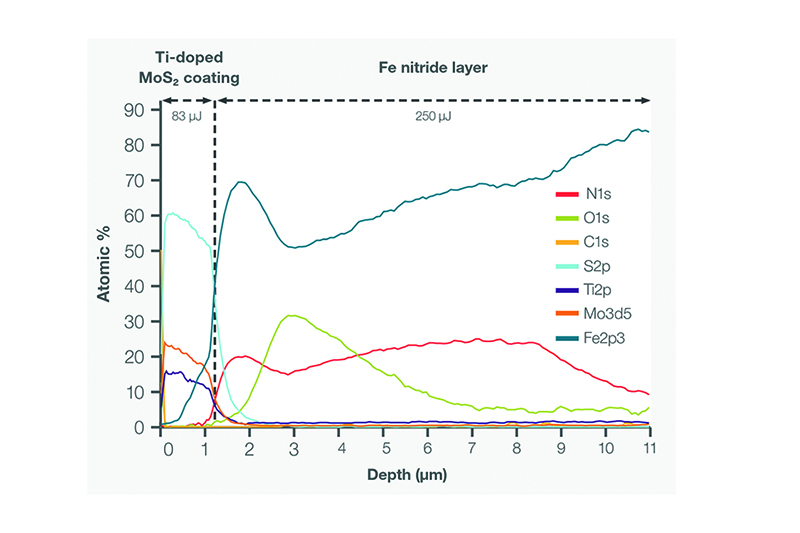
The image below shows the measurement of a nitrided steel sample with a molybdenum disulfide and titanium (MoS2:Ti) coating. The femtosecond laser ablation profile took seven hours to measure 11µm and maintained the correct sample composition during the analysis. Comparatively, an ion beam profile took 30 hours to measure 6µm and damaged the MoS2:Ti coating giving an incorrect stoichiometry in the profile.
The ability to access greater depths expands the scope of XPS. Structures that were once difficult to analyse, such as interfaces beneath thick coatings or multi-layer organic paint structures, can now be examined directly. Interfaces that lie tens of micrometres below the surface become accessible without the need for destructive sectioning techniques.
A winning combination
Despite these advantages, it is important to recognise that no single technique provides a universal solution. Monatomic ion beams are highly effective for inorganic materials, offering well-understood behaviour and high-depth resolution. Gas cluster ion beams continue to be the preferred choice for many organic and polymer systems, providing a balance between depth and chemical preservation.
Femtosecond laser ablation adds a new dimension to this landscape. It is particularly valuable where chemical integrity is critical, deeper regions need to be accessed quickly, or traditional methods introduce unacceptable artefacts.
As materials science continues to evolve, so too will the demands placed on analytical techniques. Modern materials are increasingly complex, combining multiple phases, intricate architectures and sensitive chemistries. Understanding them requires tools that are both powerful and adaptable.
The evolution of XPS depth profiling reflects this broader trend. What began as a straightforward extension of surface analysis has become a nuanced discipline, shaped by the need to balance precision, speed and chemical fidelity.
More like this...