Multi-tasking, bio-inspired endoscope could enable ‘remote surgery’
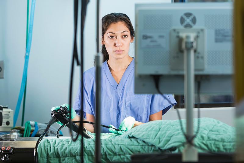
A bio-inspired medical endoscope features an optical design that combines the high-resolution 3D imaging of human vision with the mantis shrimp’s capability to simultaneously detect multiple wavelengths of light.

This could enhance identification of diseased tissues and, by adding fluorescence imaging, can make cancerous tissue light up for easier removal or highlight anatomy critical parts that need to be avoided during surgery.
Created by a group of researchers at the Chinese Academy of Sciences, the multi-modal 3D endoscope comprises three parts – a broadband binocular optical system, an optical relay system and a multi-band sensor.
The binocular optical system works similar to the human eye to obtain high-resolution 3D information. The multi-band sensor – which consists of pixels with different spectral and polarisation responses – is bio-inspired by the compound eyes of the mantis shrimp. These can detect multi-spectral information while also recognising polarised light.
‘Existing fluorescence 3D endoscopes require surgeons to switch working modes during operation to see the fluorescence images,’ explains Chenyoung Shi, one of the researchers working on the study. ‘Because our 3D endoscope can acquire visible and fluorescent 3D images simultaneously, it not only provides more visual information, but can also greatly shorten the operation time and reduce risks during surgery.
‘By introducing an optical relay system, the high-resolution 3D images after the broadband binocular optical system can be projected onto one and the same multi-band sensor. Therefore, high-resolution 3D images containing visible light and fluorescence information are recorded.’
Shi describes how ‘precision optical processing and on-chip spectroscopy technology made this multi-modal 3D endoscope possible. To obtain high-quality 3D images, the binocular optical system requires two sets of optical systems with exactly the same parameters’.
Shi adds that this places high requirements on the processing accuracy of optical components. The wider band coating ensures the simultaneous imaging of visible and near-infrared fluorescence. On the other hand, the fabrication of the bio-inspired multi-band sensor requires the integration of a wafer-level filter array on a CMOS sensor.
The shell of this endoscope is made of biocompatible 304 stainless steel, while the optical lens is made of K9 glass or SF57 glass. The lens surface is coated with silicon dioxide (SiO2) or magnesium fluoride (MgF2) to achieve 400-1,000nm anti-reflection, and the illumination fibre is made of high-purity optical glasses. The chemical-resistant glasses allow repeated cleaning and autoclaving cycles with minimum losses in transmission. During the operation, a near-infrared imaging reagent named ICG – approved by the US Food & Drug Administration (FDA) – is used to label tumour tissues.
The resulting endoscope has been designed for robotic surgery systems that help increase the precision and accuracy of minimally invasive procedures. The enhanced visual information could help surgeons in a different room from the patient clearly distinguish various types of tissue in the surgical field.
Could this pave the way for future remote surgery? ‘Remotely controlled robotic surgery requires not only real-time 5G high-speed network, but also comprehensive information on the patient’s lesion,’ Shi explains. ‘This bio-inspired multi-modal 3D endoscope will provide the information of intraoperative visualisation and location of the tumour tissue, lymph nodes and vital structures without impeding surgical workflow.’
For now, the researchers plan to use the instrument to perform additional biological and clinical imaging. They also intend to incorporate more wavelengths and the ability to sense polarisation to provide even more visual information.
Shi concludes, ‘In our next research, some met surface materials, such as plasma structure or photonic crystal material, will be used for spectral separation and polarisation detection. A large number of experiments are necessary before this system could be used clinically. If it moves through clinical trials successfully, it can directly replace the existing endoscope, [and] the doctor does not need to be familiar with this new system.’


