Implant promotes skull growth
An engineered, triphasic, biomaterial scaffold could help treat craniosynostosis.
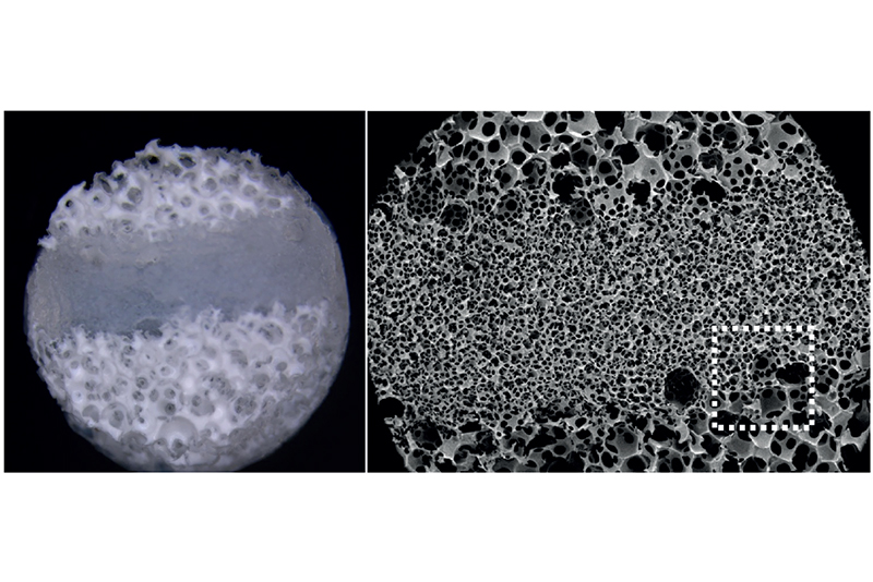
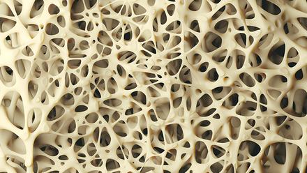
Triphasic poly-L-lactic acid scaffold showing distinct pore regions, interconnected structure and nanofibrous surface
© Adapted from Benton Swanson, W., Douglas, L., Woodbury, S.M. et al. A tissue engineering approach to regenerate the cranial suture skeletal stem cell niche with a multicompartment biomaterial scaffold. Bone Res 14, 58 (2026). https://doi.org/10.1038/s41413-026-00539-z http://creativecommons.org/licenses/by/4.0/Craniosynostosis is a condition that causes premature fusion of skull bones, restricting growth and placing pressure on the developing brain.
Rather than relying on drugs or growth factors, the approach uses controlled pore sizes to recreate the environment needed for normal skull growth.
Treating craniosynostosis usually requires surgery to cut the joint back open. Researchers in the US claim to have developed a poly-L-lactic acid (PLLA) implant that rebuilds the cranial stem cell niche the joint relies on for growth, which is lost in craniosynostosis.
The study, led by Professor Yuji Mishina at the University of Michigan with Dr Ben Swanson, now at the Harvard School of Dental Medicine, tested the scaffold in an animal model brain.
'The current standard, suturectomy and cranial vault remodelling, treats the anatomical consequence rather than the cellular cause,' Swanson explains.
'Reopening a fused suture leaves that depleted niche unaddressed, which is why re-synostosis is common, revision surgery frequent, reported surgical complication rates run as high as 22.9%, and no pharmacological option exists.'
A natural suture consists of stem-cell-rich fibrous tissue bordered by two actively forming bone surfaces. To replicate this ‘bone-suture-bone’ architecture, the team designed a three-part PLLA scaffold with a central, small-pore, stem cell reservoir and larger-pored compartments on either side to promote bone formation and blood vessel growth.
Previous tissue engineering approaches have largely sought to suppress bone formation, such as by altering signalling or limiting blood vessel growth. Those approaches 'create a soft-tissue gap but do not reconstitute a functional, host-integrated, stem cell niche', says Swanson. 'Our construct instead recreates the bone-suture-bone unit in a single vascularised, host-integrating implant.'
The scaffold was made using sugar spheres to set the pore sizes and thermally induced phase separation to create a nanofibrous surface.
The resulting 50-500nm fibres mimic a collagen-scale structure, promoting protein adsorption and cell adhesion. Scanning electron microscopy confirms distinct but connected pore regions, allowing cells, nutrients and blood vessels to move through the implant.
Previous work showed that very small pores encourage stem cells to remain immature – partly by suppressing the mechanical signals that would otherwise push them to form bone – while larger pores promote bone-forming cells and blood vessel growth.
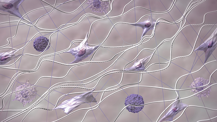
Using fluorescently labelled stem cells, the researchers found that differentiating cells migrated from the small-pore centre into larger-pore regions, while stem cells that resemble the inner cell mass of a pre-implantation embryo are concentrated in the centre.
'[Pore size] governs not only whether cells differentiate but whether they migrate – and that differential migration is the sorting mechanism that keeps stem cells central and osteogenesis peripheral,' says Swanson.
Even under unusually strong bone-forming signals, the central compartment resisted bone formation and stayed rich in stem cells. 'We did not assume that pore geometry alone would hold the central compartment stem-like…yet it did,' says Swanson. 'That argues that the mechanical microenvironment can override a strong biochemical differentiation cue.'
The scaffold, in turn, maintains open and suture-like tissue, and stopped the joint fusing again. The animal models show normal skull growth, with the biggest improvements when the implant was placed earlier in development.
Although the results are encouraging, Swanson emphasises that the work remains an early proof-of-concept. 'The principal hurdles are cell sourcing, scale, long-term safety and regulatory path,' he says. Reproducing the scaffold’s sharp, interconnected multi-zone structure under manufacturing-grade conditions is a challenge.
Further research must also show that normal skull growth continues after the scaffold degrades and establish the best treatment window in human infants.
Swanson believes the same principle could be applied to other tissues where stem cells sit between two tissue types, including tendon-to-bone attachments, cartilage and periodontal tissue.