Bone grown in lab to test medical treatment
A bone-on-a-chip device could offer an alternative to animal research for trialling treatments of diseased or damaged bone.
‘This study mainly focuses on the development of necessary 3D architecture and mechanical stimulation suitable for bone tissue engineering, drug development and long-term culture,’ says Dr Frederik Claeyssens, Reader in Biomaterials at the University.
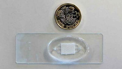
In collaboration with Universitat Ramon Llull, Spain, the team has manufactured a 3D microfluidic device that is suitable for short and long-term experiments. The technology consists of a microfluidic chamber, a 3D scaffold made of polymerised high internal phase emulsion (polyHIPE), and a fluidic pump that controls the mechanical stimulation and facilitates the exchange of nutrients and metabolic wastes. Emulsion testing has been used to engineer the 3D structure with the polyHIPE material.
The polyHIPE is made using an oil that solidifies under ultraviolet light to create a plastic material made up of millions of tiny, interconnected holes where the water used to be. These highly porous materials form a scaffold that helps cells create new bone tissue in 3D.
The team has also investigated an appropriate mechanical stimulation for osteogenesis – also known as brittle bone disease – that mimics the human body’s dynamic environment.
‘These developments will lead…to accurately investigating novel therapeutics for bone and sequestering of toxins into bone, in comparison to standard in vitro and in vivo testing, which could lead to a breakthrough in the field of osteogenesis,’ Claeyssens notes.
‘For bone tissue engineering, besides biocompatibility, scaffolds should have a highly porous structure and a suitable mechanical stability. This highly porous HIPE structure with appropriate pore size for cells to fit in (10-50 microns diameter) allows enough room to accommodate cells of varying 3D shapes and, through smaller interconnecting pores, allows them to freely interact with the neighbouring cells in a 3D environment similar to bone in vivo conditions. This also enhances nutrient diffusion and metabolic waste removal.’
As the device hosts microenvironments that are similar to 3D in vivo conditions, the team claims it can be used as a powerful substrate to investigate bone mechanobiology. Claeyssens adds that computational fluid dynamics can be added to investigate the effects of shear stress factors on bone cells in a 3D substrate.
‘Microfluidic technology can provide high-throughput assays for identifying suitable drug concentrations and combinations,’ Claeyssens says. ‘Importance of microfluidic developments is their ability for drug screening and delivery at the cellular level in vitro. The technology allows precise control of drug flow to the culture chamber, allowing the researcher to real time monitor and study cellular responses.’
Claeyssens states that the current model has been designed, fabricated and successfully assessed, showing potential to be used as a platform for bone pharmaceutical studies. ‘However, further studies are needed to improve the model, investigate bone physiology and test different therapeutic compounds.’