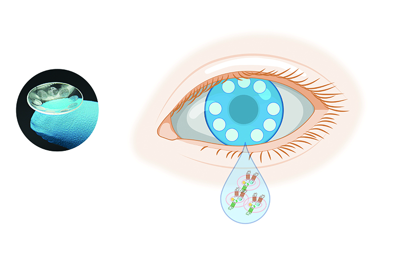
A contact lens to detect disease
The biosensors in the smart lens could improve cancer diagnosis, prognosis and treatment.
Historically, researchers have applied conventional methods to detect exosomes – extracellular vesicles – as potential biomarkers for cancer diagnosis because they play an important role in the metastasis of cancers. These detection methods include immune-based assay and microRNA analyses, but isolating the nanometre-sized vesicles in sufficient quantity and purity presents a challenge.
According to scientists at the Terasaki Institute for Biomedical Innovation (TIBI) in Los Angeles, USA, current methods involve 'tedious and time-consuming ultracentrifuge and density gradients', which can take at least 10 hours to complete. In addition, the most common detection methods are expensive and require large equipment.
'We are the first team to use a contact lens with the integration of the colorimetric-based bio-sensing assay to capture and detect exosomes,' says Assistant Professor Yangzhi Zhu.
'In addition, our reported contact lens biosensors have excellent optical transparency and mechanical properties similar to commercial contact lenses.'
Although other bodily fluids such as blood, urine and saliva contain substantial concentrations of exosomes, tears do not contain high levels of cellular debris, water or fat-soluble metabolite and protease, which interfere with disease diagnosis. As Zhu notes, the contact lens offers “an ideal interfaced device to the eye or tear” and could provide helpful medical information for various diseases.
With this in mind, the scientists have designed an anti-body-conjugated signalling micro-chamber contact lens or ABSM-CL to capture the exosomes contained in tears. Using nanoparticle-tagged specific antibodies to stain the ABSM-CL, the scientists report that they can visually detect and select the exosomes.
As part of the device’s preparation, the researchers use a direct laser cutting and engraving approach to fabricate the micro-chambers for the lens. This differs from the conventional method of cast moulding for the structural retention of the chambers and the lens.
The scientists have also introduced a method that chemically modifies the micro-chamber, activating its surfaces to capture the antibodies that detect the exosomes.
Zhu explains that in conventional approaches, researchers have to integrate precious metallic or carbon nanomaterials into the contact lens’ structure to immobilise the antibody. However, this process requires a clean-room facility and drives up fabrication costs.
In contrast, the team modifies its surface techniques for the polymer contact lens by applying organosiloxane molecules.
'The [silicon-oxygen] Si-O backbone of the organosiloxane molecule can form stable covalent bonding with any surface presented with [hydroxide] OH moieties,' explains Zhu. 'Since the polymer surface is enriched in OH groups, this approach bypasses the need to incorporate metallic elements into the contact lenses. In addition, integrating reactive amine groups on the organosiloxane molecule allows antibodies to attach onto the contact lenses with greater ease.'
A gold nanoparticle-based colorimetric assay is used to visualise the captured exosomes spectroscopically.